Post an image of a light or electron microscope specimen that was imaged and processed digitally. Include a description of the specimen, microscopy type, and magnification.
All posted evidence
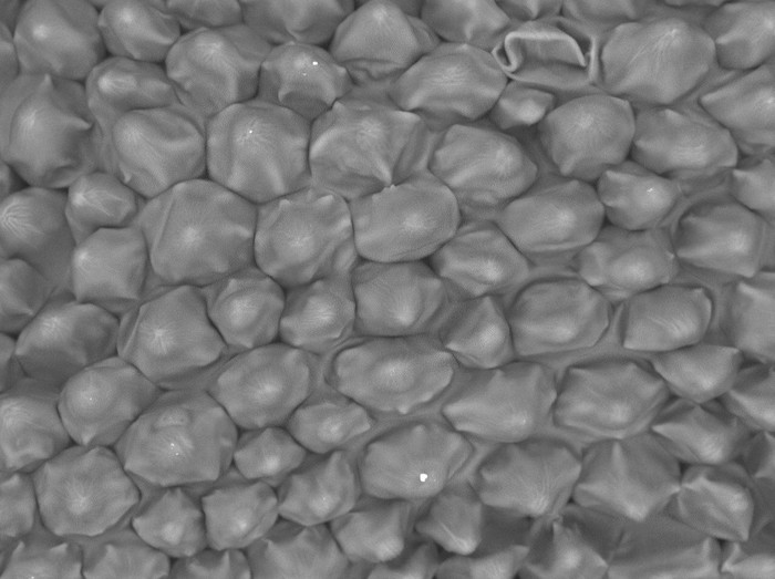
Orchid Central Part (x180) SEM- Eden Mercado
edenm
Over 9 years ago
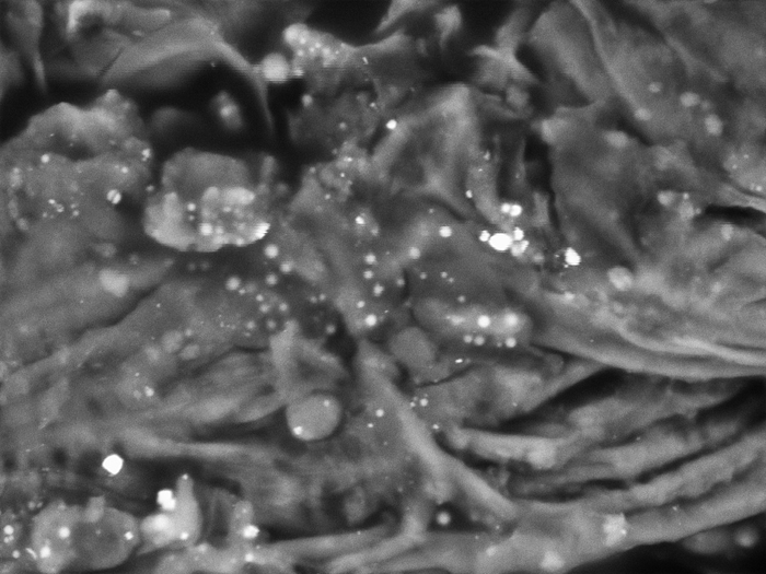
Posted Image
colemeier24
Over 9 years ago
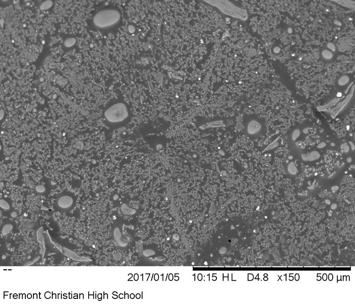
Cheddar Cheese 140x
vincentfcs
Over 9 years ago
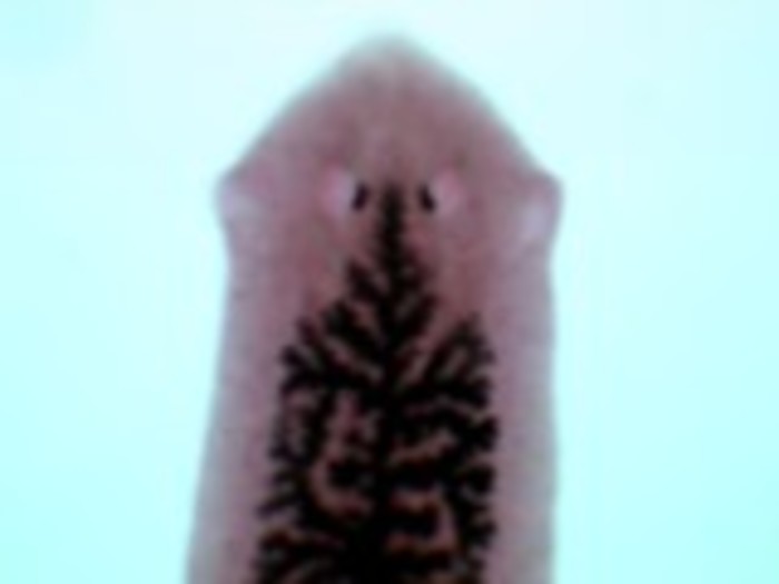
leaf from photoshop.
There was a problem processing this image, the file may be of an invalid format.
Uploaded Filename:
johnny leaf new 4x .tif
johnny
Over 9 years ago
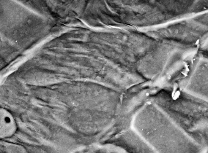
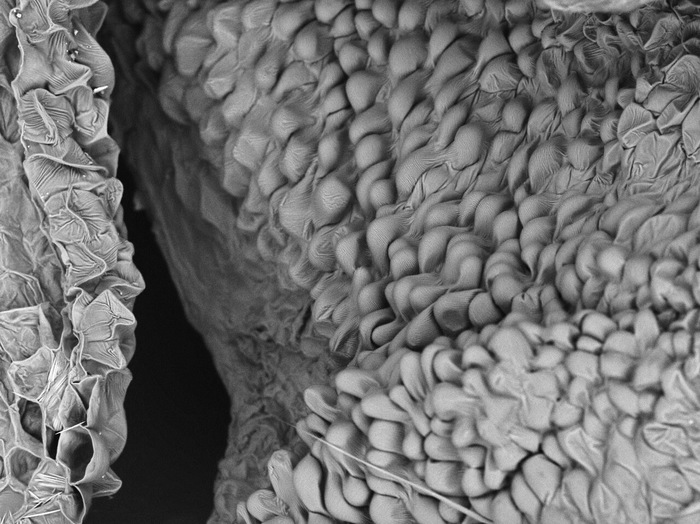
Garlic, x1800 , Electron Microscope
julianaabraham
Over 9 years ago
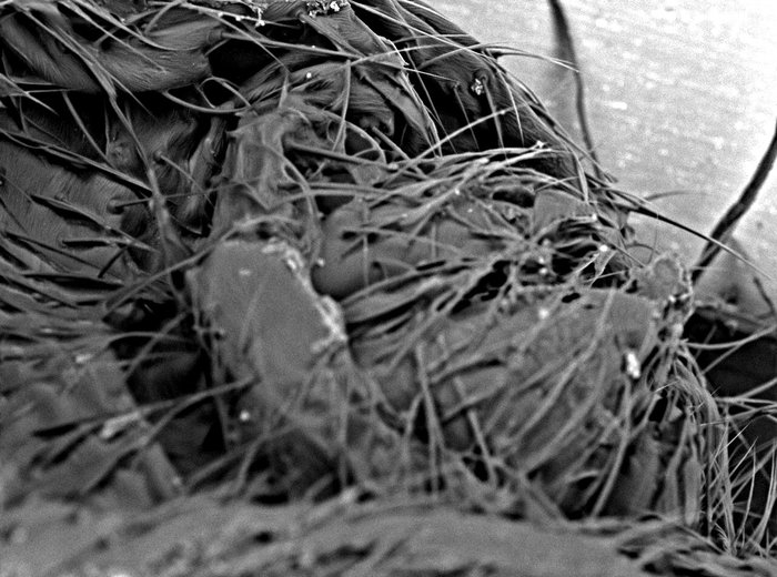
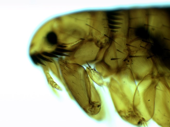
End part of Spider's Head. 200x Magnification. Electron Microscope

 johnny
Over 9 years ago
johnny
Over 9 years ago