Sign up
Sign in
Introduction-to-Electron-and-
/
Digital-Image-Processing-in-Microscopy
/
Digital Microscopy Image 1
Digital Image Processing in Microscopy
Digital Microscopy Image 1
Only editable by group admins
Last updated December 30, 2016 at 2:24 PM
Evidence visible to public
Post an image of a light or electron microscope specimen that was imaged and processed digitally. Include a description of the specimen, microscopy type, and magnification.
All posted evidence
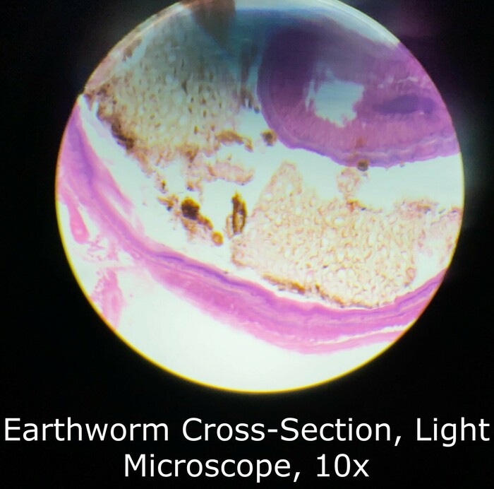
Earthworm Cross-Section, Light Microscope, 10x Magnification
micah
Over 4 years ago
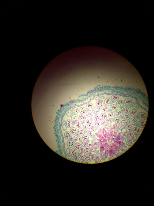
Frog Testes Light Microscope x4-Lawrence. pixlr
lawrence45
Over 8 years ago
paramecium bursaria conjugation chromosomes 10x
xuwilliam
Over 8 years ago
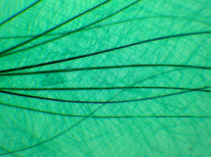
Down feather 40x
inmanmax
Over 8 years ago
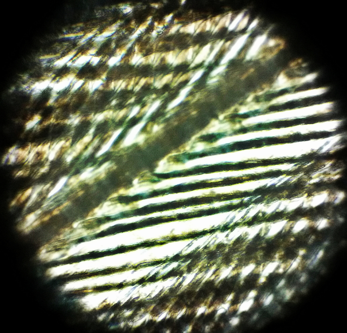
Feather x400 Light Microscope
jay_c
Over 8 years ago
Endamoeba histolytica sec. Z116 40X
tony666
Over 8 years ago
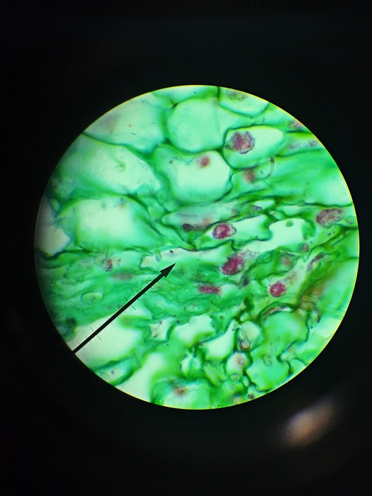
frog blood, 100x
alice-z
Over 8 years ago
Penicillium Cincidiophores sec. of an Orange Peel , 100x
breanna3
Over 8 years ago
Odigonium 40x
maranda123
Over 8 years ago
oscillatoria wm 40x
jerryhuang
Over 8 years ago
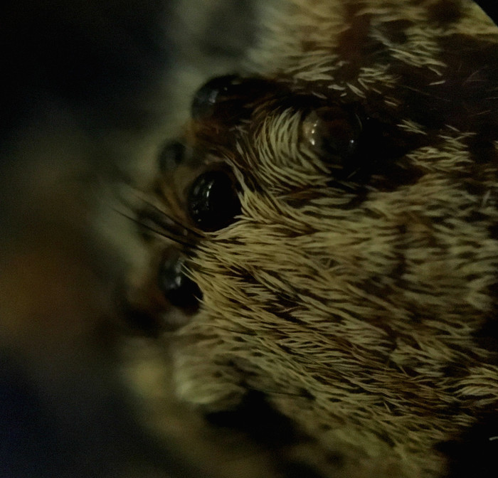
Spider under Stereoscope, Edited, 60x
rachelhandran
Over 8 years ago
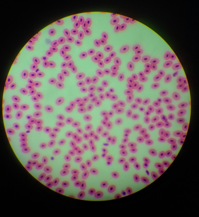
Human Blood x400
sonalisidhu
Over 8 years ago
New version available
Refresh
Dismiss