Post an image of a light or electron microscope specimen that was imaged and processed digitally. Include a description of the specimen, microscopy type, and magnification.
All posted evidence
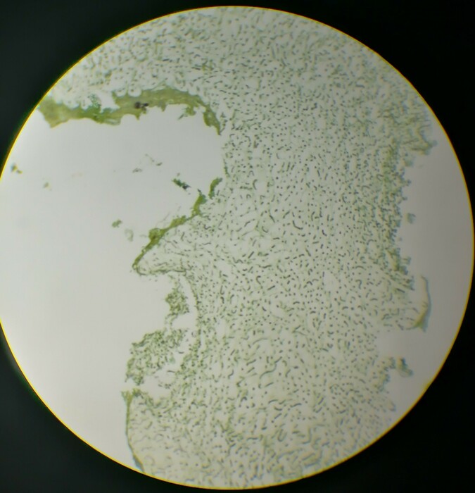
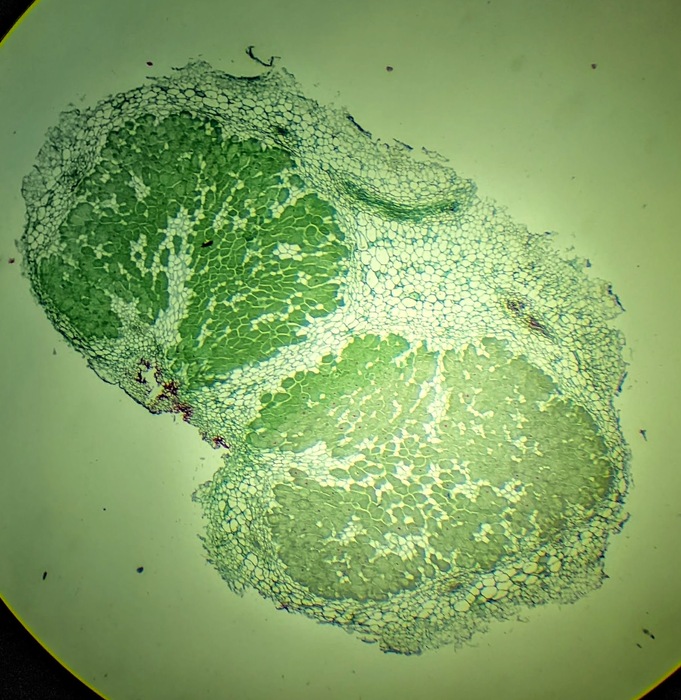
Nostoc Section of Nodule
austinmo
Over 4 years ago
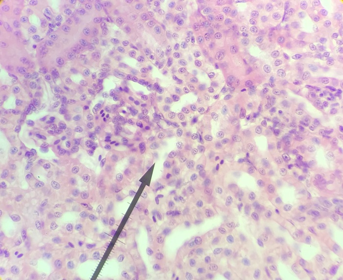
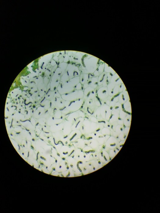
Frog Kidney 40x
alexandria3
Over 4 years ago
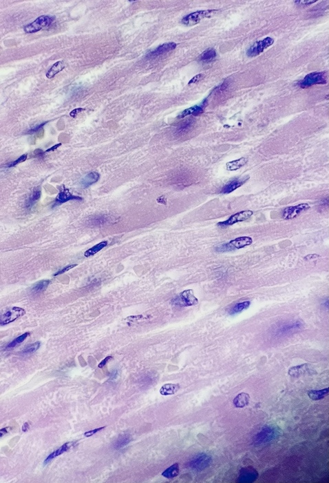
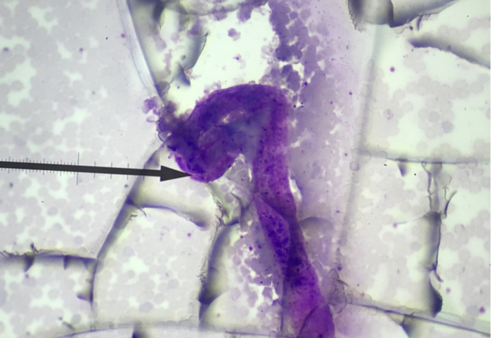
Cardiac muscle
Microscope
10x
emma-hebert
Over 4 years ago
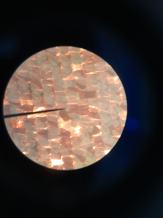
feather. 10X. use light microscope
jessie-zhang
Over 4 years ago
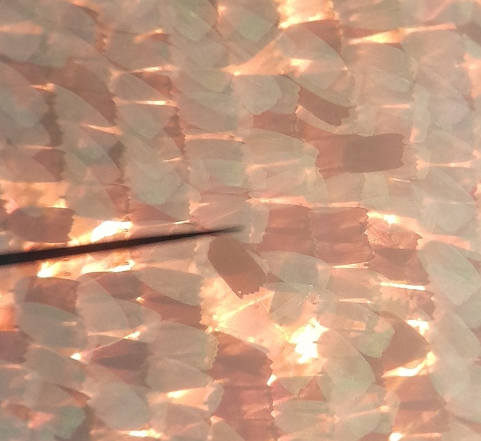
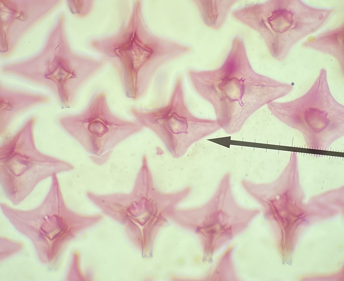
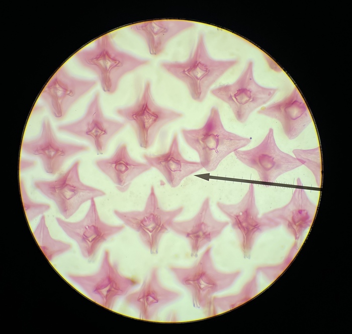
Fish scale placoid w.m., Light Microscope, 40x
cjin002
Over 4 years ago
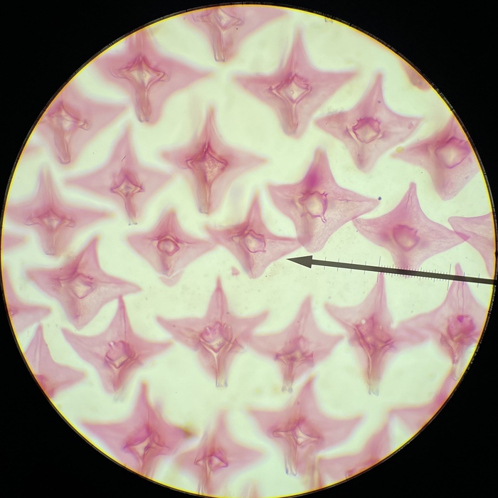
Fish scale placoid w.m., Light Microscope, 40x
cjin002
Over 4 years ago
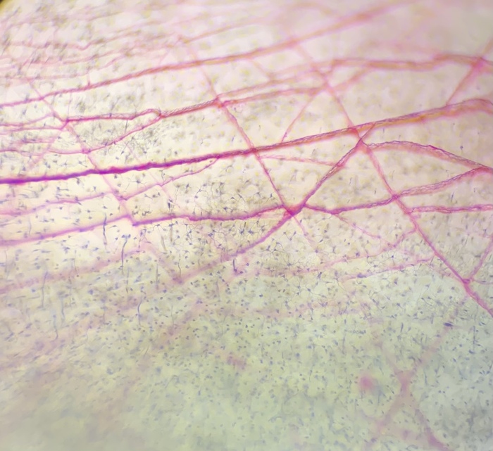
Fish scale ganoid wm- loose connective tissue- light microscope 10x