Digital Image Processing in Microscopy

Digital Microscopy Image 4

Only editable by group admins

  • Last updated December 30, 2016 at 2:24 PM by drhandran
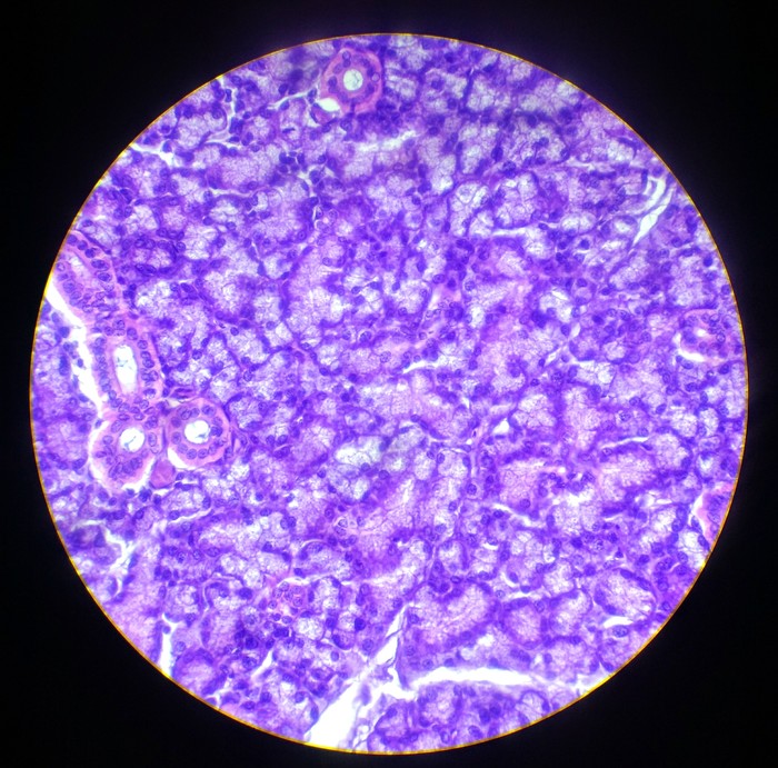
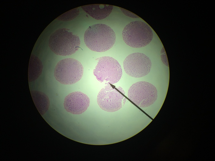
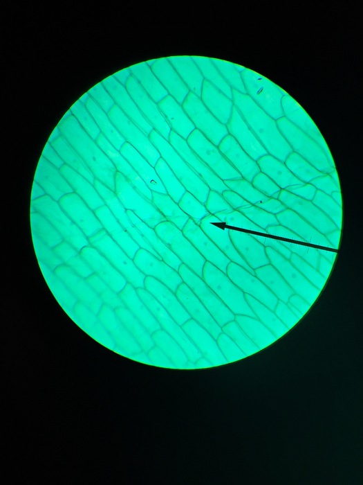
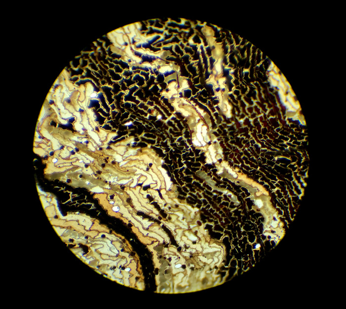
  • Evidence visible to public
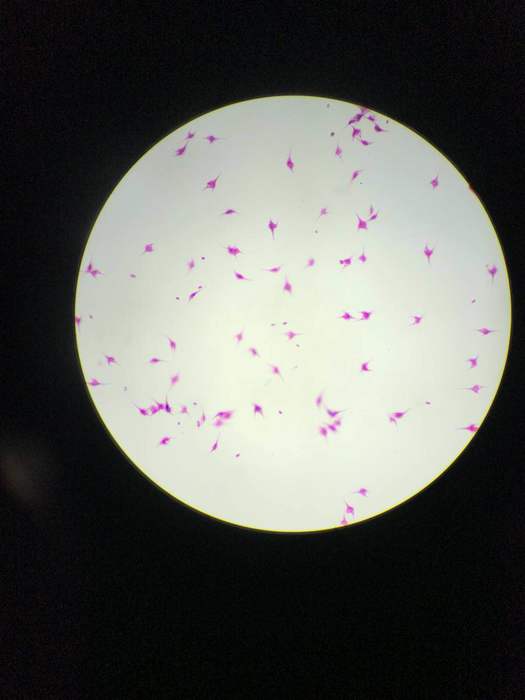
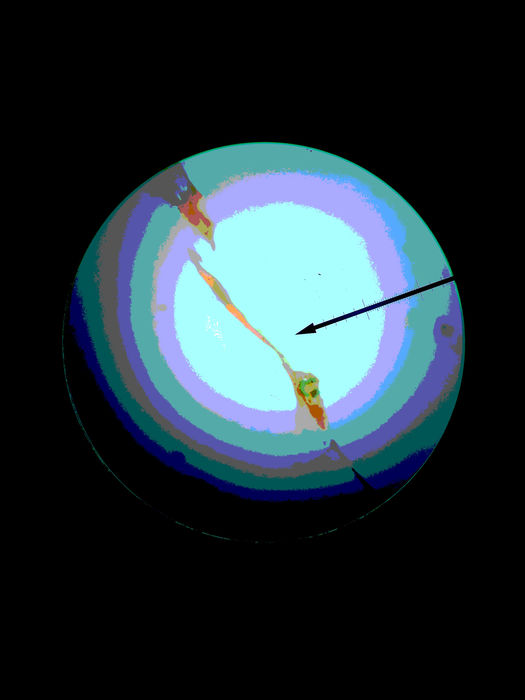
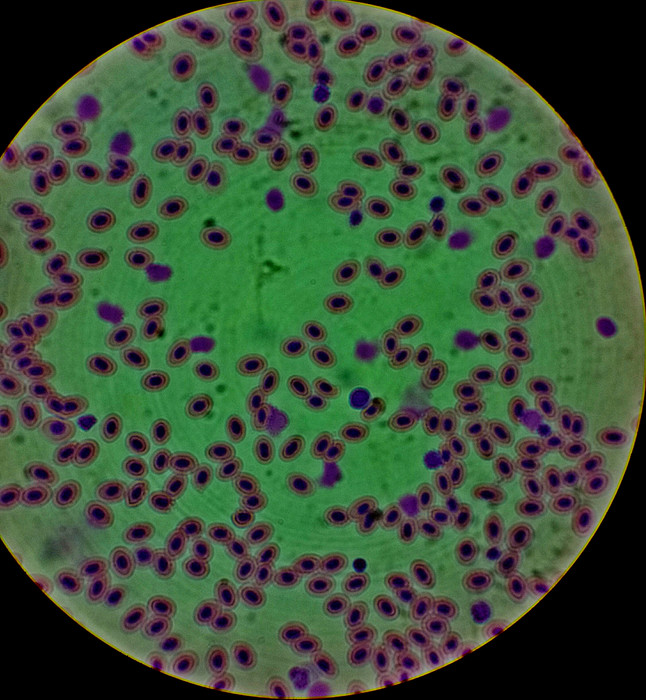
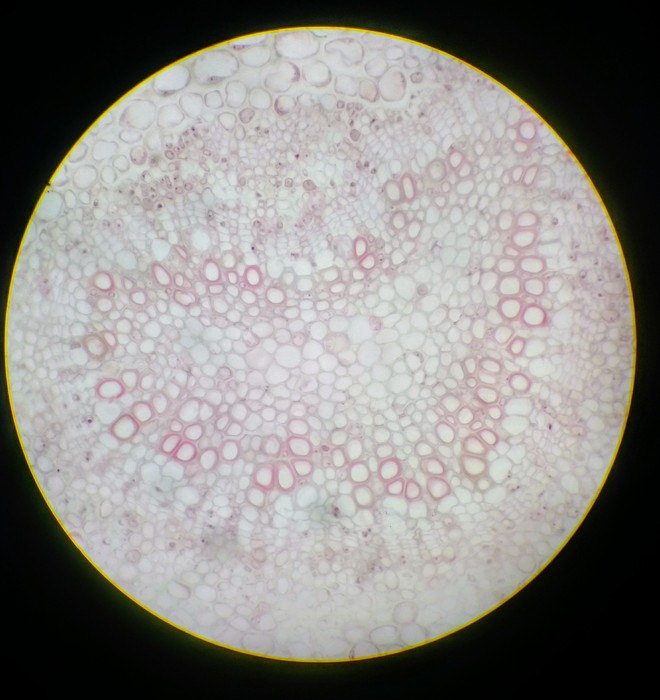
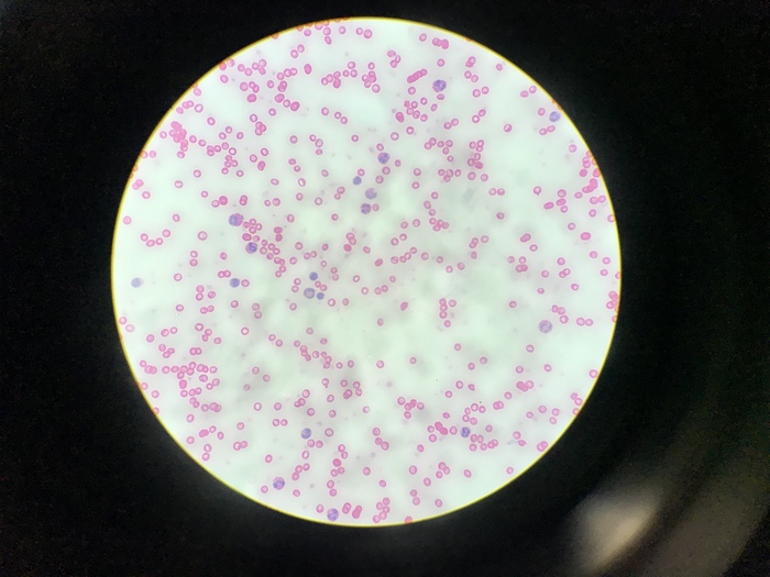
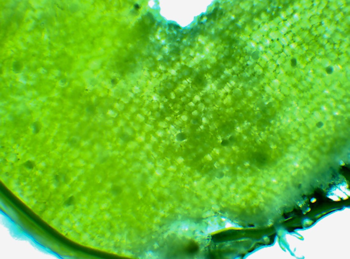
Post an image of a light or electron microscope specimen that was imaged and processed digitally. Include a description of the specimen, microscopy type, and magnification.

All posted evidence

ce ra tiu m 10x

xuwilliam Almost 8 years ago

Human Smear Light Microscope x4-Lawrence pixlr

lawrence45 Almost 8 years ago

Frog Blood x400 Light Microscope

jay_c Almost 8 years ago

Syringa leaf, 400x

alice-z Almost 8 years ago

Salivary gland mammal, submaxillary 400x

inmanmax Almost 8 years ago

Zee kernel & embargo I.s 40X

tony666 Almost 8 years ago

Mitochondria (wm) af&f Allium Epidermal , 10x

breanna3 Almost 8 years ago

coccus 100x

maranda123 Almost 8 years ago

Pine Cone Scale, Edited, 100x

rachelhandran Almost 8 years ago

dirofilaria immitis x400

jerryhuang Almost 8 years ago

Human Chromosome Debris Female Smear x400

sonalisidhu Almost 8 years ago

Orchid Stamen (40x) Light Microscope

edenm Almost 9 years ago