Last updated December 30, 2016 at 2:24 PM
by drhandran
Evidence visible to public
Post an image of a light or electron microscope specimen that was imaged and processed digitally. Include a description of the specimen, microscopy type, and magnification.
All posted evidence
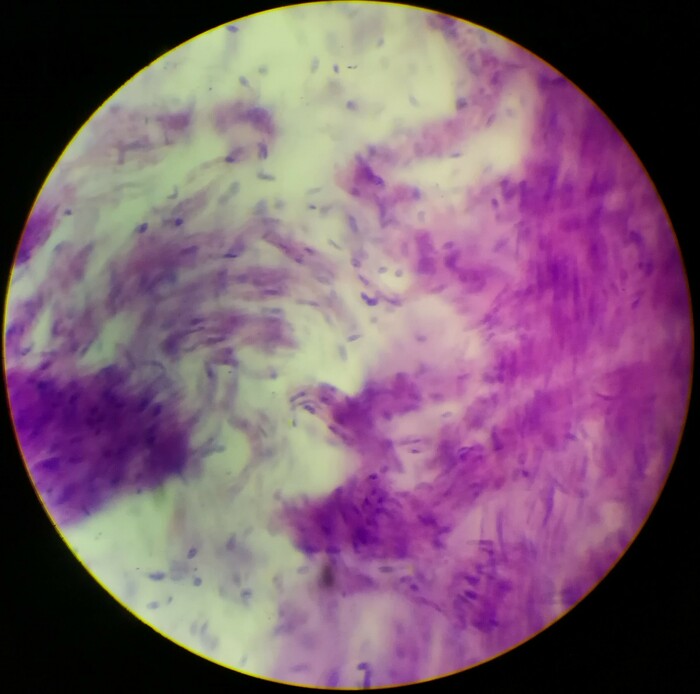
Spleen 40x
austinmo
Over 4 years ago
Shark embryo
Microscope
40x
emma-hebert
Over 4 years ago
Bone, developing long Ls. 40X use light microscope
jessie-zhang
Over 4 years ago
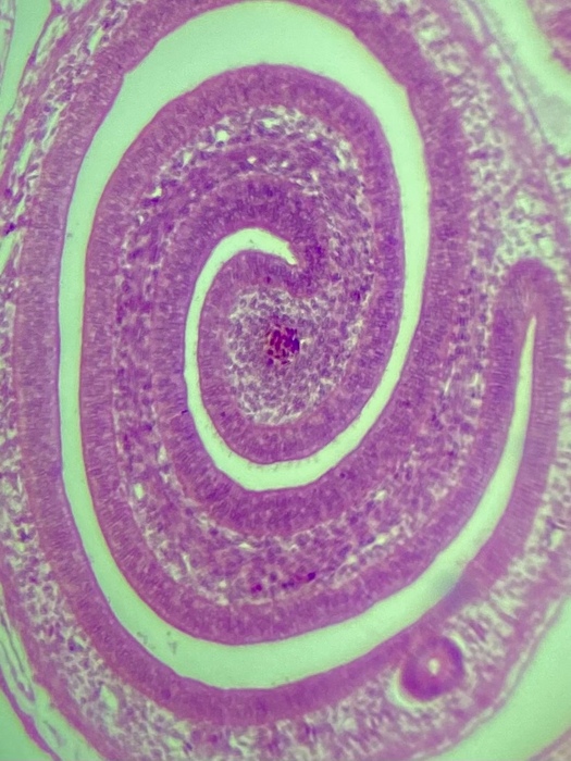
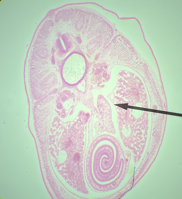
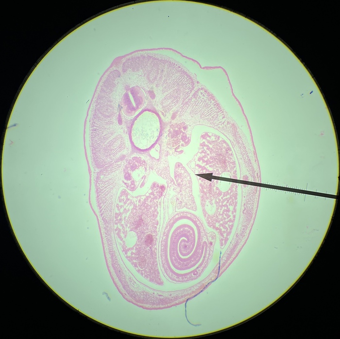
Shark Embryo 50mm, Light Microscope, 4x
cjin002
Over 4 years ago
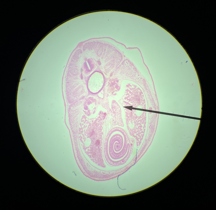
Shark Embryo 50mm, Light Microscope, 4x
cjin002
Over 4 years ago
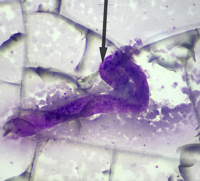
Shark Embryo 50mm, Light Microscope, 4x
cjin002
Over 4 years ago
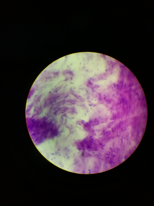
Plasma 40x
alexandria3
Over 4 years ago
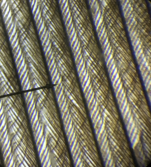
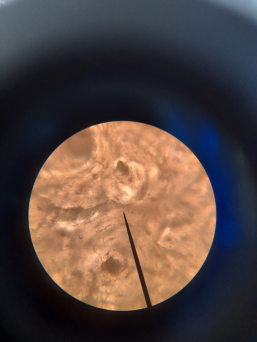
Aves feather
light microscope
10x
catherine-zhang
Over 4 years ago
Tensed Voluntary Muscle Piece
10x Zoom
shanmukh
Over 4 years ago
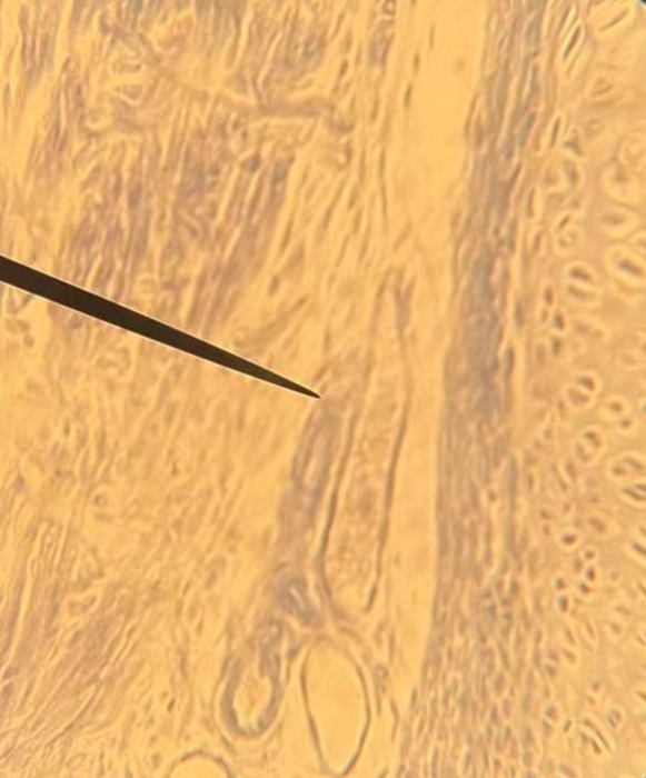
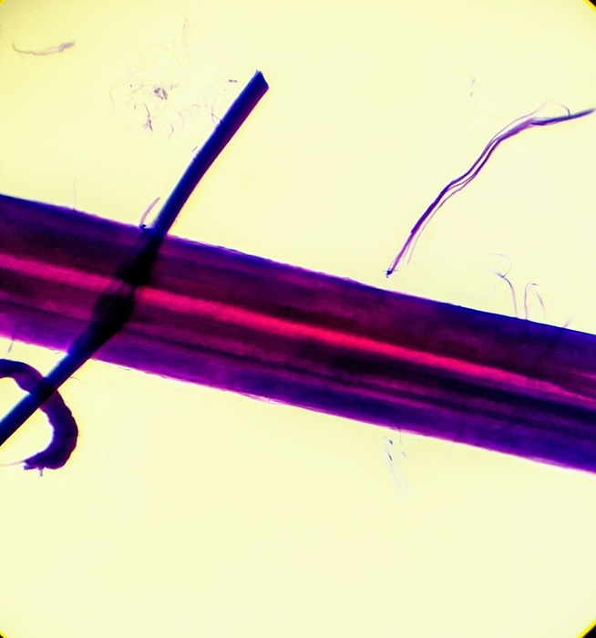
Posted Image
austinmo
Over 4 years ago
Ground bone, cs. Human. 10X. use light microscope.