Sign up
Sign in
Introduction-to-Electron-and-
/
Scanning-Electron-Microscopy
/
Electron Microscopy Image 3
Scanning Electron Microscopy
Electron Microscopy Image 3
Only editable by group admins
Last updated May 9, 2018 at 6:50 PM
Evidence visible to public
Post an image of an electron microscopy specimen that was collected, prepared, and imaged on the Hitachi TM3000 by you. Include a description of the specimen and magnification.
All posted evidence
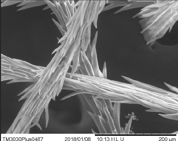
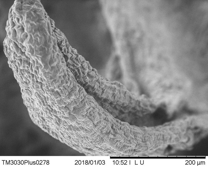
hair 2000x
jerryhuang
Over 8 years ago
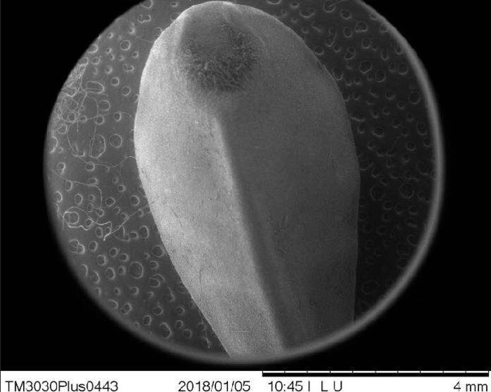
seed 1000
xuwilliam
Over 8 years ago
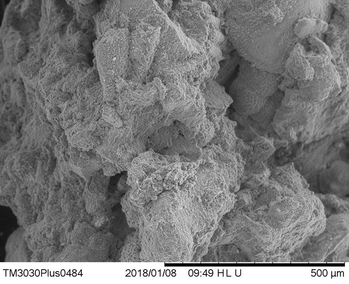
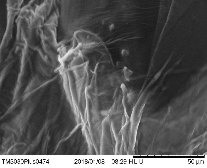
Bark 2500x
inmanmax
Over 8 years ago
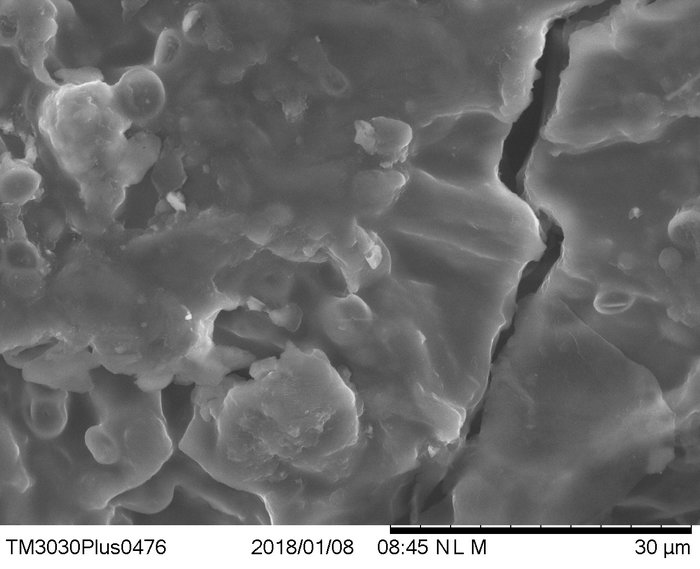
Sand , 180x
breanna3
Over 8 years ago
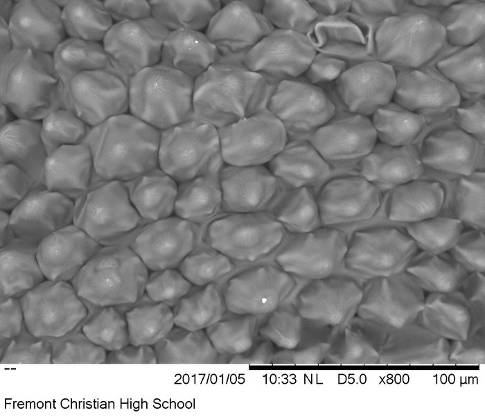
Seedling x400
jay_c
Over 8 years ago
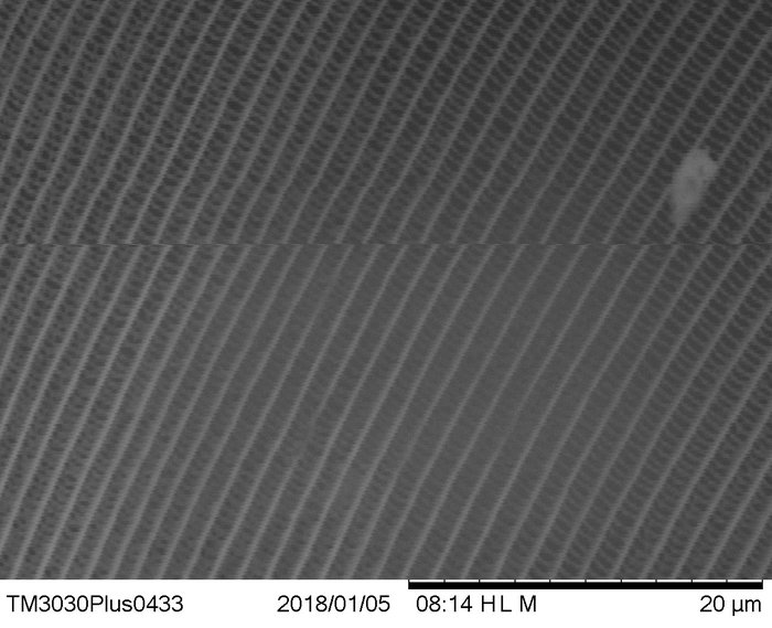
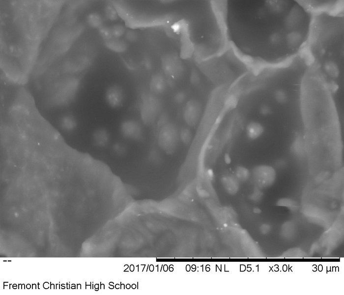
White Onion x1200
sonalisidhu
Over 8 years ago
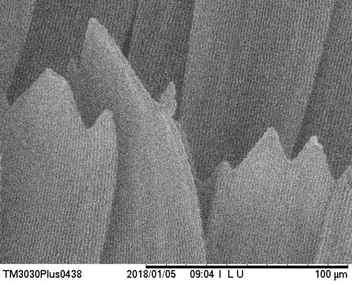
Butterfly Wing.x4000-Lawrence Shou
lawrence45
Over 8 years ago
butterfly scale, 1000x
alice-z
Over 8 years ago
Snake Skin, 3000x
rachelhandran
Over 8 years ago
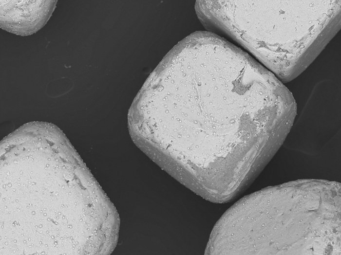
Iodized salt cubes. 150x magnification
solomon
Over 9 years ago
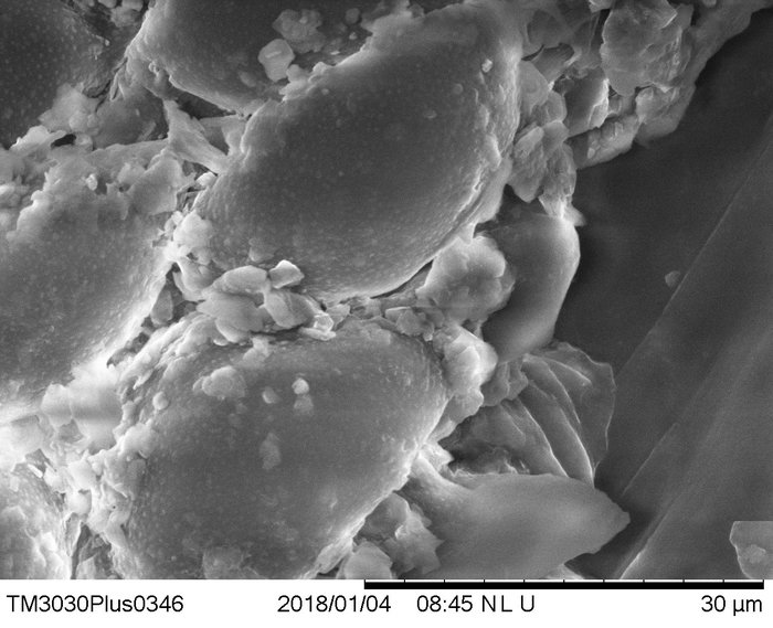
almond slice 3,000x
vincentfcs
Over 9 years ago
Red Flower 800x Magnification
ashna-ghuman
Over 9 years ago
New version available
Refresh
Dismiss