Sign up
Sign in
Introduction-to-Electron-and-
/
Scanning-Electron-Microscopy
/
Electron Microscopy Image 2
Scanning Electron Microscopy
Electron Microscopy Image 2
Only editable by group admins
Last updated May 9, 2018 at 6:50 PM
Evidence visible to public
Post an image of an electron microscopy specimen that was collected, prepared, and imaged on the Hitachi TM3000 by you. Include a description of the specimen and magnification.
All posted evidence
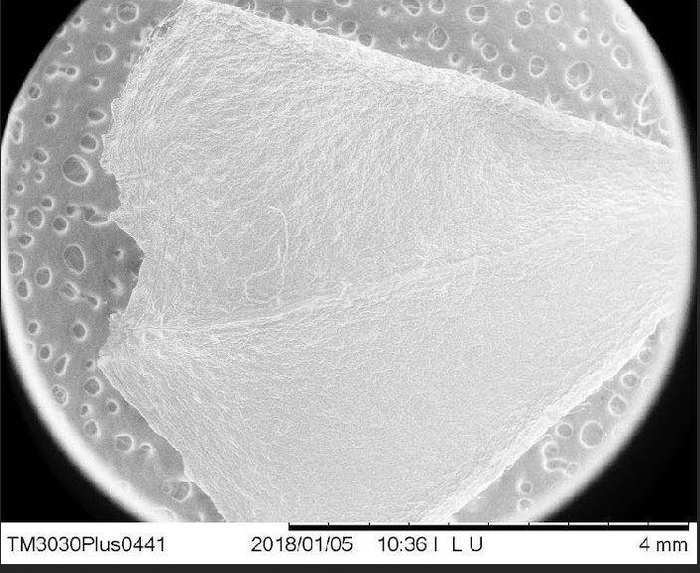
Leaf 1000
xuwilliam
Over 8 years ago
seed 1000x
jerryhuang
Over 8 years ago
Pencil shavings? 400x
inmanmax
Over 8 years ago
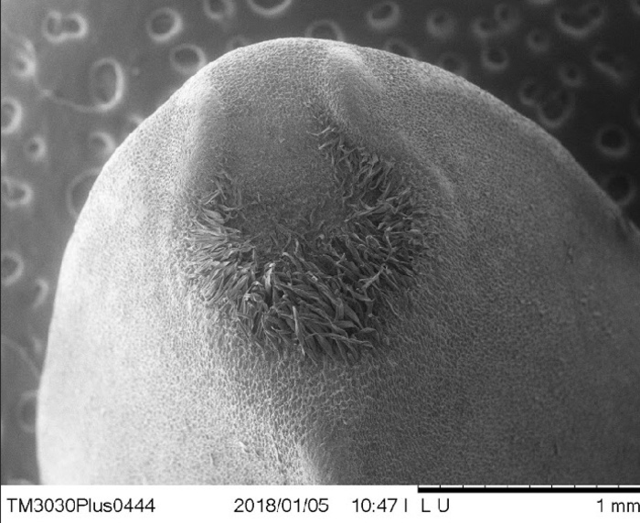
Flower part 1 , 40x
breanna3
Over 8 years ago
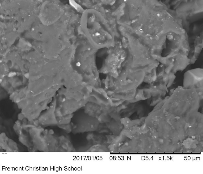
Dust x1500
jay_c
Over 8 years ago
White Onion x1200
sonalisidhu
Over 8 years ago
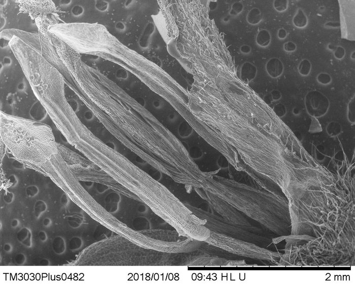
Plant Seed.x1200-Lawrence Shou
lawrence45
Over 8 years ago
seed root, 800x
alice-z
Over 8 years ago
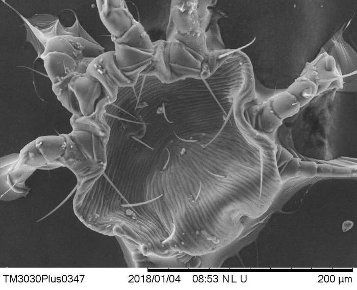
Dust Mite, 500x
rachelhandran
Over 8 years ago
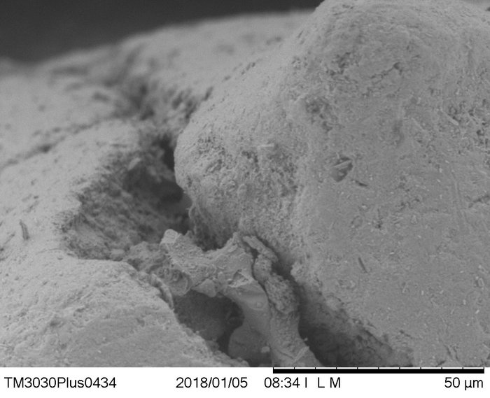
egg shell 1,800x
vincentfcs
Over 9 years ago
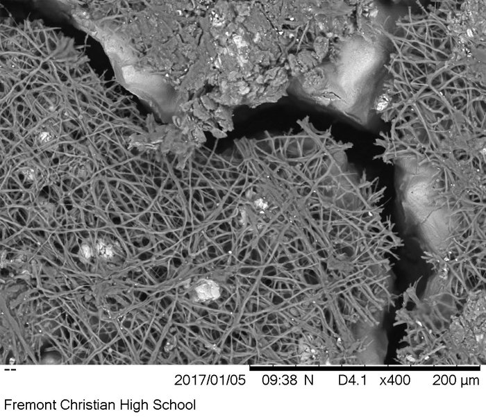
scent stick x1.5k , 1500 magnificaton
ashna-ghuman
Over 9 years ago
egg shell 400x
colemeier24
Over 9 years ago
New version available
Refresh
Dismiss