Post an image of a light or electron microscope specimen that was imaged and processed digitally. Include a description of the specimen, microscopy type, and magnification.
All posted evidence
Posted Image
kaylaj2022
Over 4 years ago
Posted Image
thedrummindude
Over 4 years ago
Posted Image
thedrummindude
Over 4 years ago
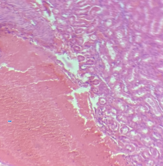
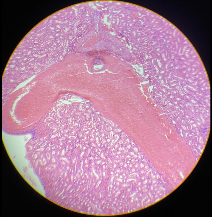
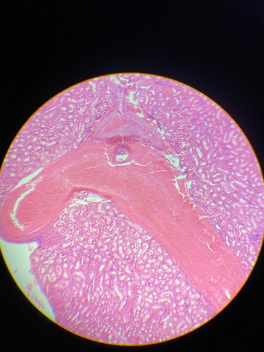
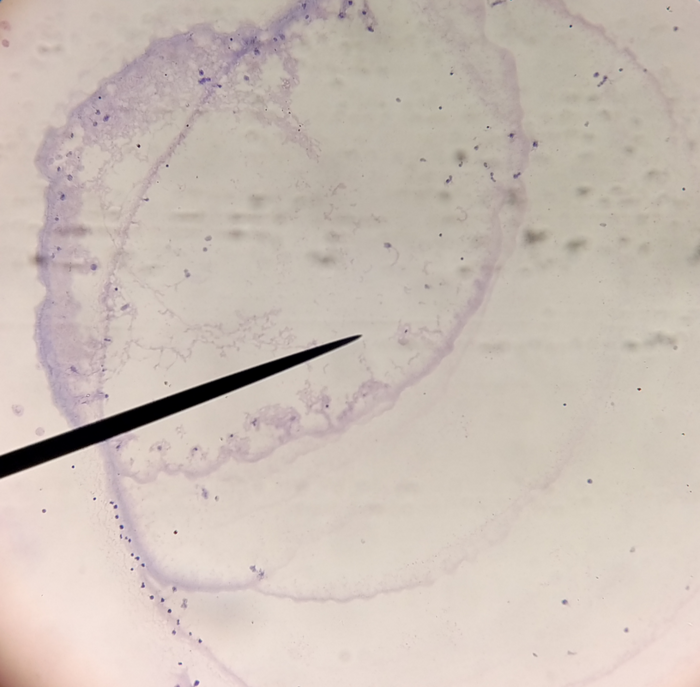
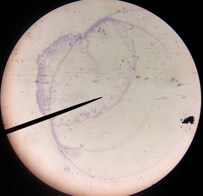
Kidney non-median sag. sec, light microscope with 4x objective.
thedrummindude
Over 4 years ago
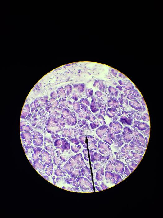
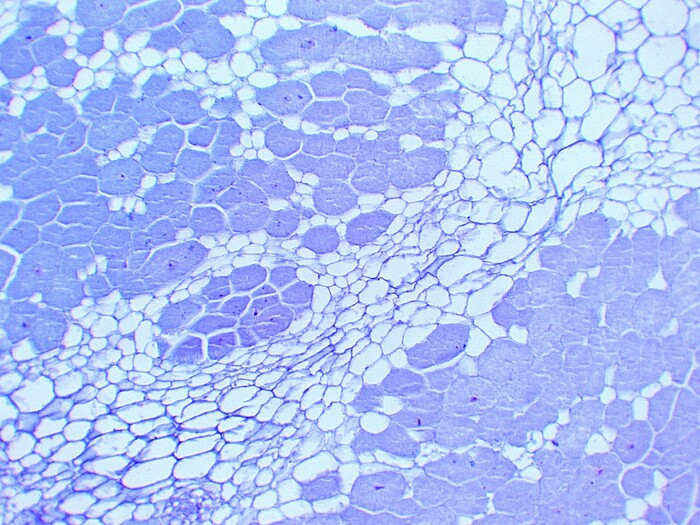
Human pancreas sec, 40x
jonathanw
Over 4 years ago
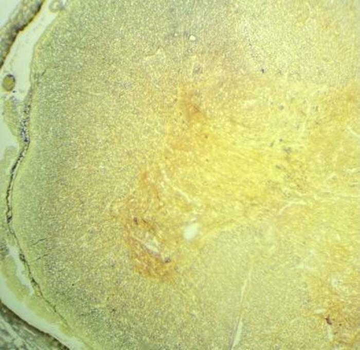
This is a image of a Spinal cord with a silver impregnation viewed under a 10x scope. This image has been edited through photos a program which is automatically installed on a windows pc.
gabem
Over 4 years ago
Female Chromosome, Light Microscope, 40x
matthew-nguyen
Over 4 years ago
Female Chromosome, Light Microscope, 40x
matthew-nguyen
Over 4 years ago
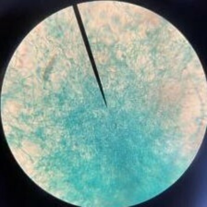
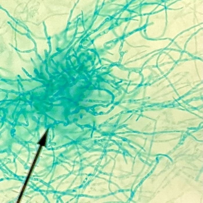
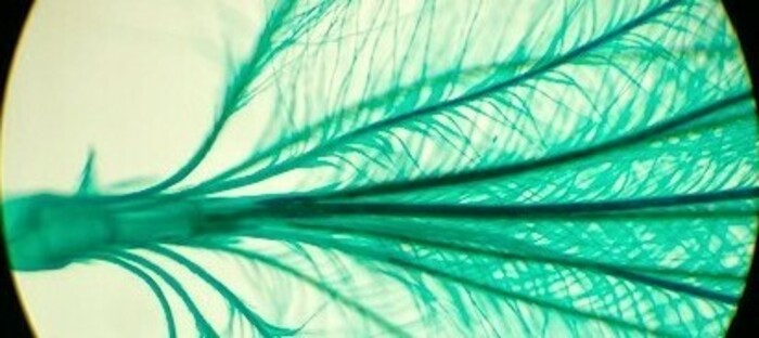
This is a image of penicillium mycelium conidiophores viewed through a 40x scope. This image has also been edited through the photos program on a windows computer.

 gabem
Over 4 years ago
gabem
Over 4 years ago