Post an image of a light or electron microscope specimen that was imaged and processed digitally. Include a description of the specimen, microscopy type, and magnification.
All posted evidence
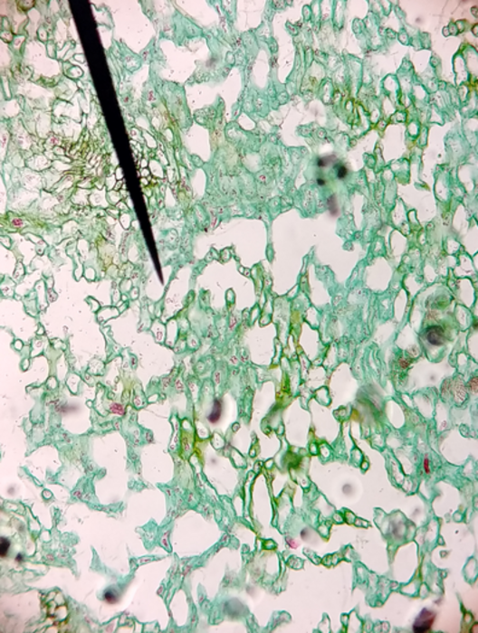
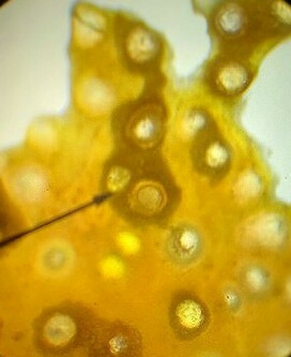
Posted Image
kaylaj2022
Over 4 years ago
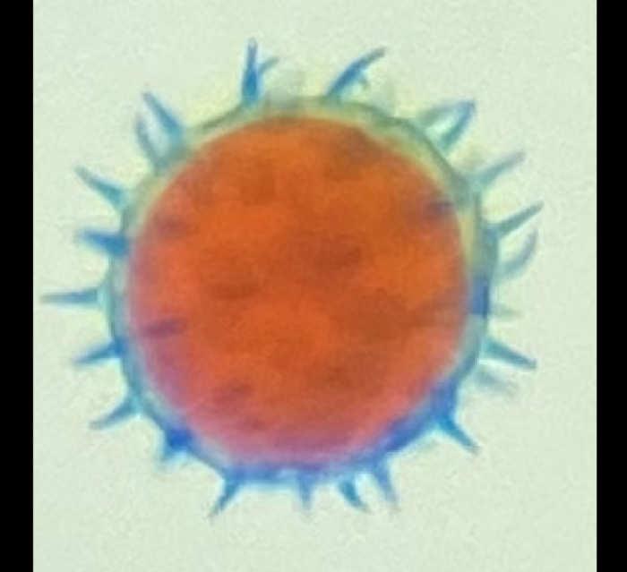
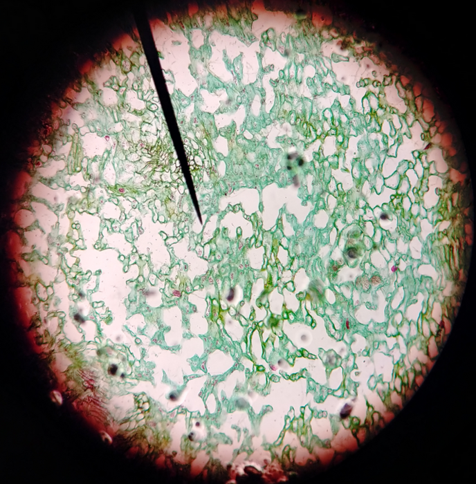
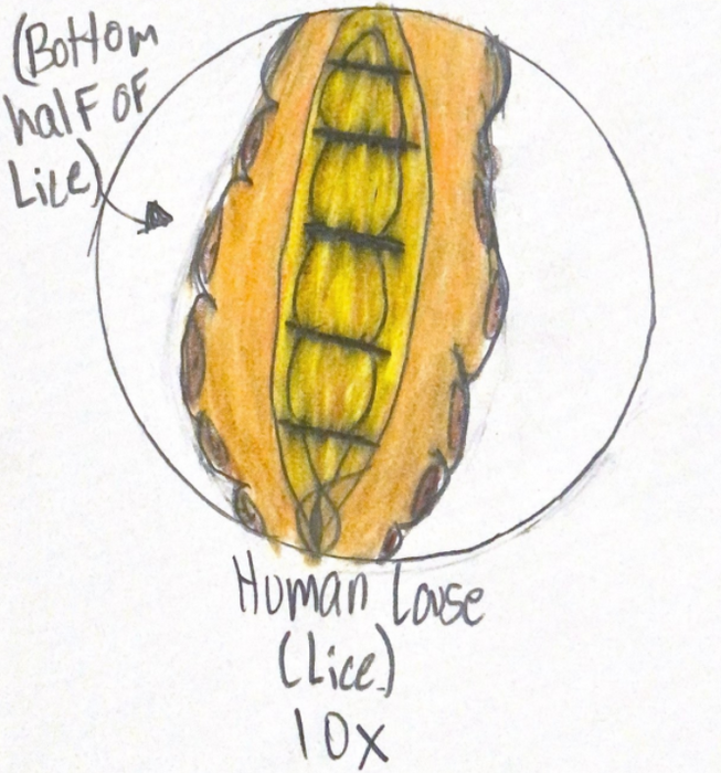
Posted Image
thedrummindude
Over 4 years ago
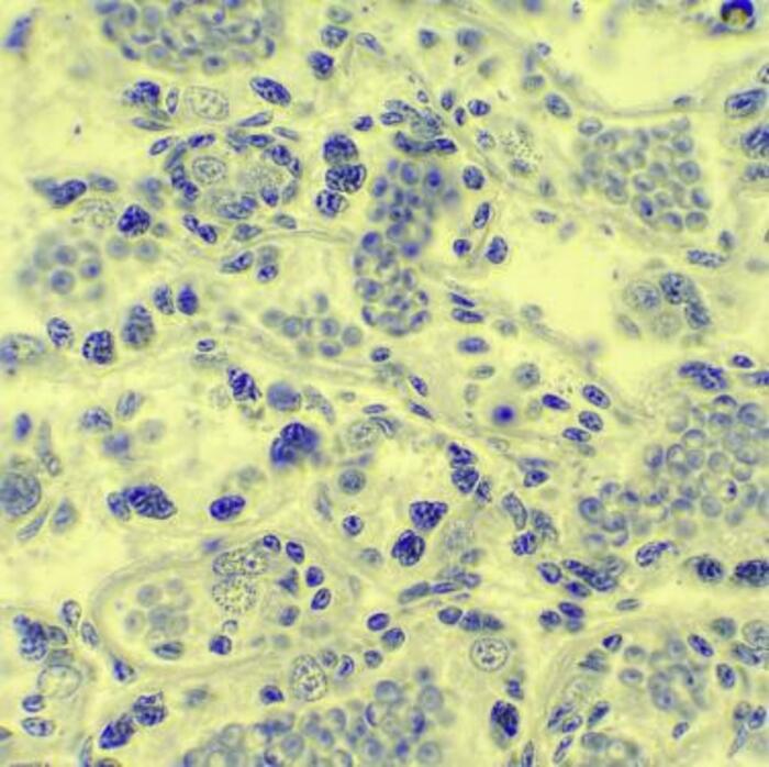
Posted Image
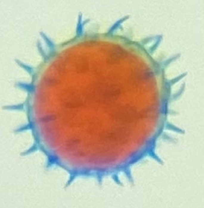
thedrummindude
Over 4 years ago
A Pollen sample under a light microscope with a 40x objective
thedrummindude
Over 4 years ago
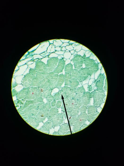
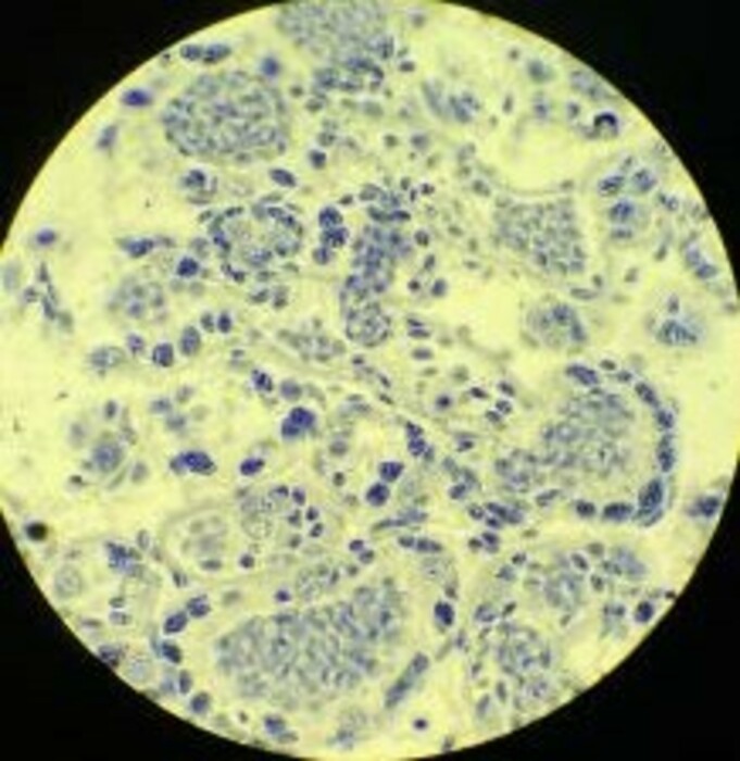
Root Tubercle legume sec., 40x
jonathanw
Over 4 years ago
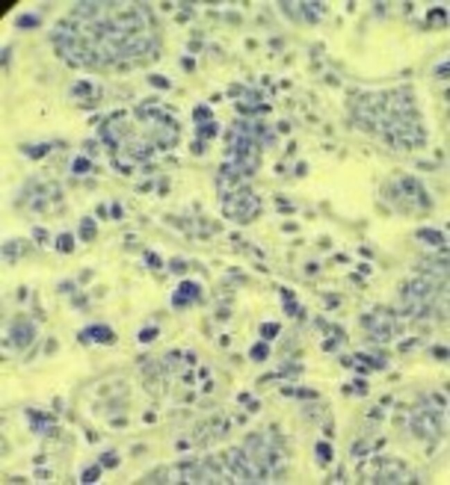
This is a picture of Crayfish Testis viewed through a 40x scope. This image has been edited through the photos program on a windows pc.
gabem
Over 4 years ago
Posted Image
gabem
Over 4 years ago
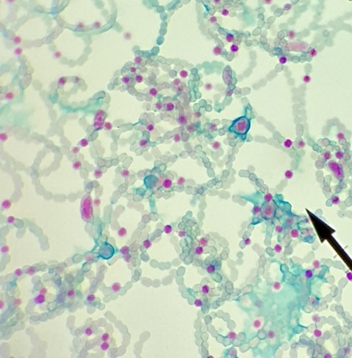
Penicillium, Light Microscope, 10x
matthew-nguyen
Over 4 years ago
Penicillium, Light Microscope, 10x
matthew-nguyen
Over 4 years ago
This is a picture of Crayfish Testis viewed through a 40x scope. This image has been edited through the photos program on a windows pc.

 gabem
Over 4 years ago
gabem
Over 4 years ago